


 下载产品说明书
 用小程序,查商品更便捷


 收藏
收藏
 对比
对比 咨询
咨询
Specificity/Sensitivity
参考图片
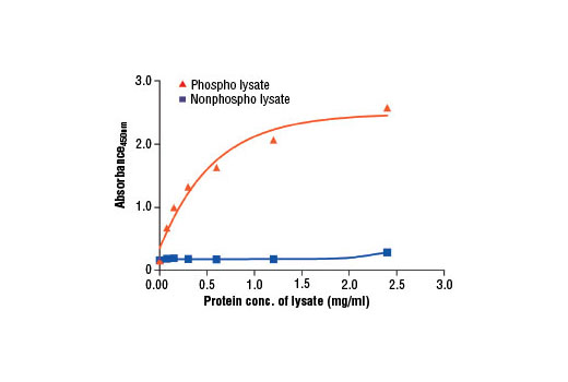
Figure 2. The relationship between protein concentration of phospho or nonphospho lysates and the absorbance at 450 nm is shown. Unstarved K-562 cells were cultured (106 cells/ml) and lysed with or without addition of phosphatase inhibitors to the lysis buffer (phospho or nonphospho lysate, respectively).图2. 磷酸化及未磷酸化的胞裂解液蛋白浓度和450nm处吸光度的关系如图所示。未饥饿处理的K-562细胞培养(106 cells/ml)且被裂解,加入或不加入磷酸化抑制剂(磷酸化或磷酸化)。
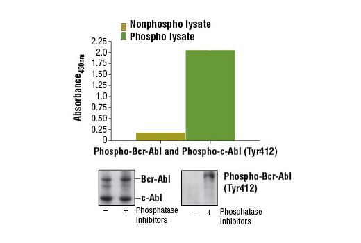
Figure 1. Constitutive phosphorylation of Bcr-Abl and c-Abl in K-562 cells lysed in the presence of phosphatase inhibitors* (phospho lysate) is detected by PathScan® Phospho-c-Abl (Tyr412) Sandwich ELISA Kit #12070. In contrast, a low level of phospho-Bcr-Abl and phospho-c-Abl protein is detected in K-562 cells lysed in the absence of phosphatase inhibitors* (nonphospho lysate). Absorbance at 450 nm is shown in the top figure while corresponding western blots using c-Abl Antibody #2862 (left panel) and Phospho-c-Abl (Tyr412) (247C7) Rabbit mAb #2865 (right panel) are shown in the bottom figure. *Phosphatase inhibitors include sodium pyrophosphate, β-glycerophosphate, and Na3VO4.图1. K-562细胞裂解后持续磷酸化的Bcr-Abl和c-Abl在phosphatase inhibitors* (phospho lysate)存在的条件下,使用PathScan® Phospho-c-Abl (Tyr412) Sandwich ELISA Kit #12070进行检测。相对的,K-562细胞中低水平的phospho-Bcr-Abl和phospho-c-Abl在phosphatase inhibitors* (nonphospho lysate)存在的条件下也能被检测到。上图中展示了450nm处的吸光值,使用c-Abl抗体#2862(左)和Phospho-c-Abl (Tyr412) (247C7)兔mAb#2865(右)进行western blot分析的结果在下图中。

 危险品化学品经营许可证(不带存储) 许可证编号:沪(杨)应急管危经许[2022]202944(QY)
  营业执照(三证合一)